
Endodontic consultation
RVG imaging or CBCT scan analysis, examination of the tooth and surrounding tissue under the microscope, temporary filling placed.
Microscope Root Canal Treatment
Specialist root canal treatment under a dental microscope — first-time and retreatment cases, including those considered difficult or hopeless elsewhere.
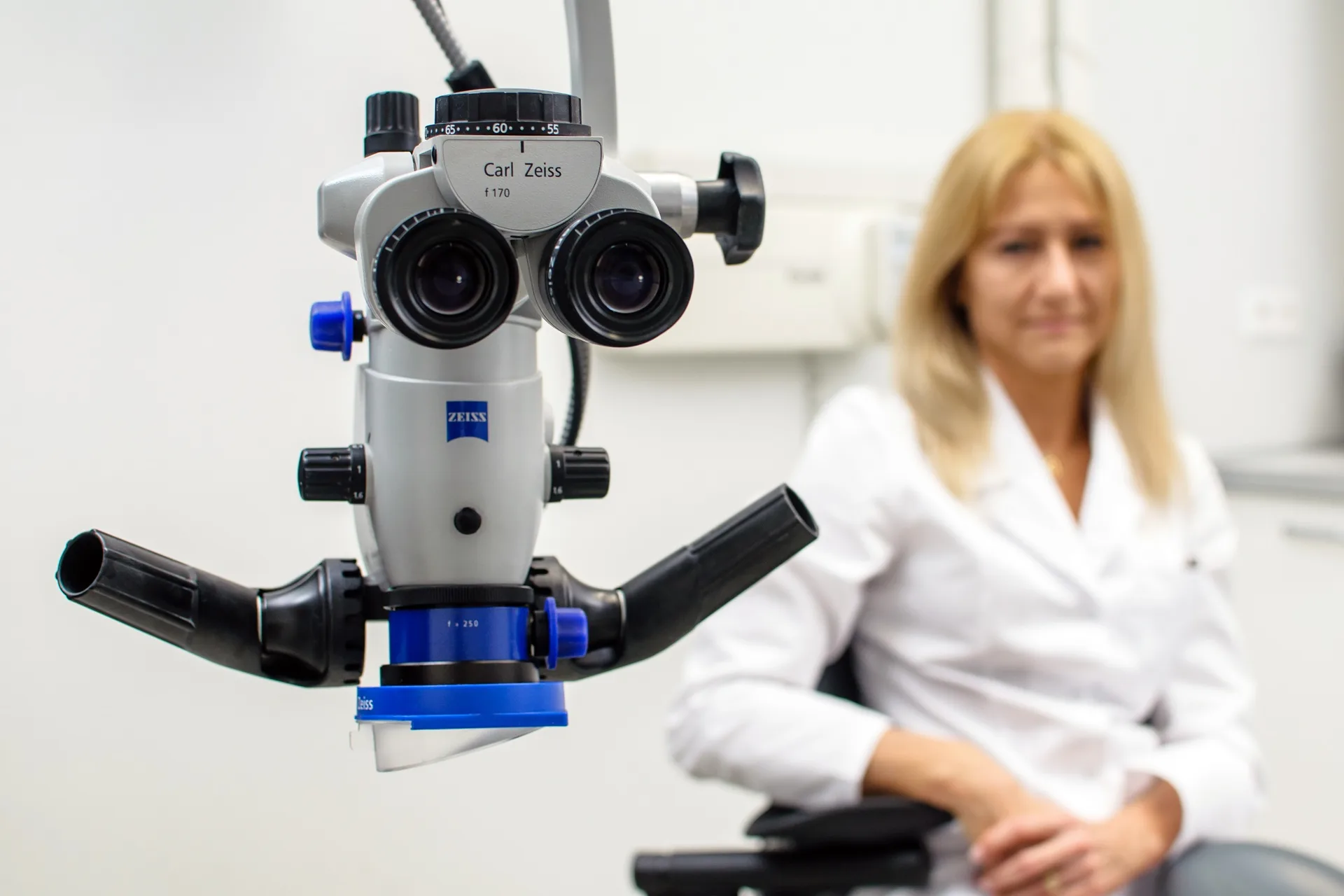
The Carl Zeiss Extaro 300 microscope shows the inside of a tooth at 8–21× magnification. This makes it possible to locate extra canals, cracks, calcifications, and other structures that simply remain invisible in traditional, unmagnified treatment — and these invisible details are most often exactly what causes a prior root canal to fail.
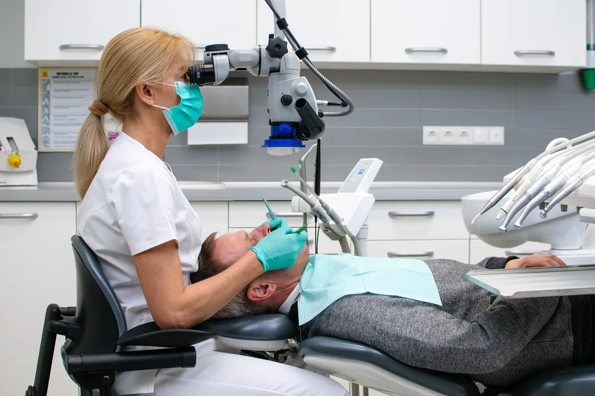
First-time root canal treatment, particularly with complex root anatomy
Retreatment after a previously failed procedure
Teeth with severely curved, calcified, or hard-to-reach canals
Cases where another dentist proposed extraction as the only option
Removing a broken instrument left in a canal from earlier treatment
Closing a perforation or an open root apex
RVG imaging or CBCT scan analysis, examination of the tooth and surrounding tissue under the microscope, temporary filling placed.
Performed under local anesthesia, typically 1.5–2.5 hours depending on the number of canals and case complexity.
A surcharge for complexity (a severely curved canal, an unusually long root, difficult access, an obliterated canal) is agreed transparently before treatment begins, not after.
An interventional visit is available if pain or swelling occurs during treatment.
A general dentist can safely perform straightforward root canal treatment. But complex cases — curved canals, retreatment, a broken instrument stuck in a canal — need both the equipment (microscope, digital diagnostics) and experience genuinely focused on endodontics alone. Dr. Alicja Dziedzicka has specialized in this area since 2008 and is a member of the Polish and European Societies of Endodontology.
Book an Endodontic Consultation
Selected examples of microscope-assisted treatment — from separated instrument removal through perforation repairs to anatomically complex retreatment cases.
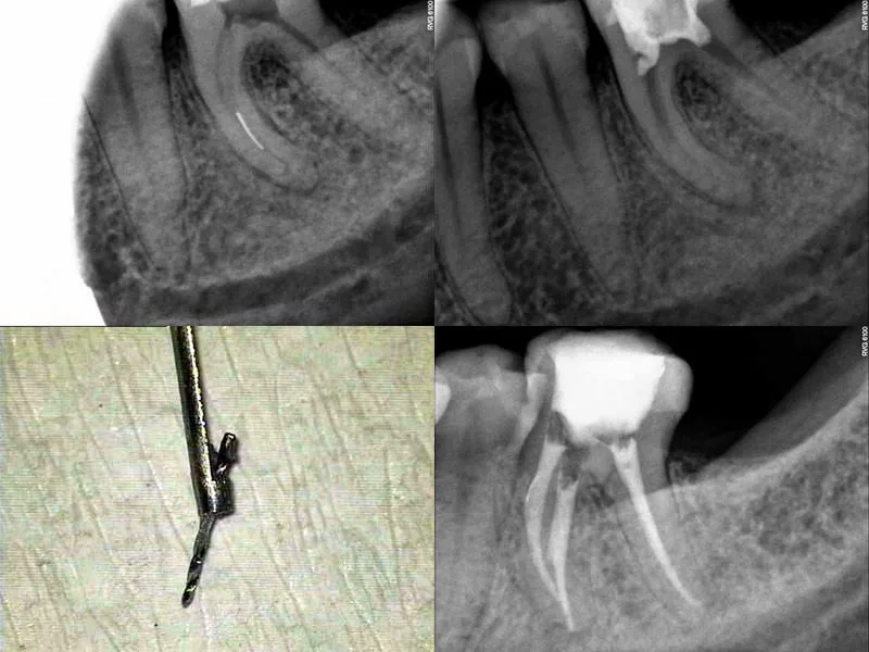 01Tooth 36
Broken instrument removal from the canal
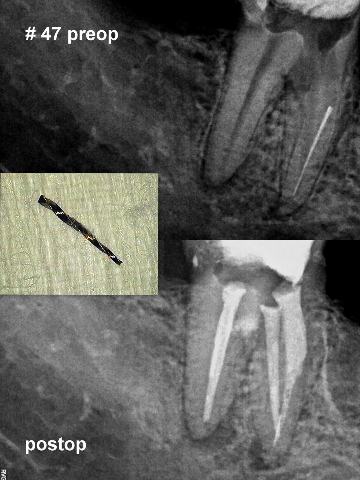 02Tooth 47
Broken instrument removal from the canal
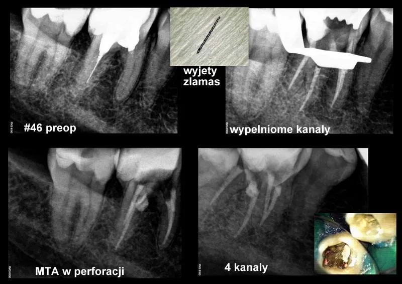 03
03Broken instrument removal and MTA perforation closure
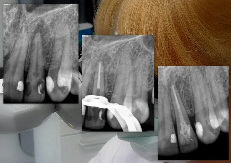 04Tooth 22
Internal resorption treatment
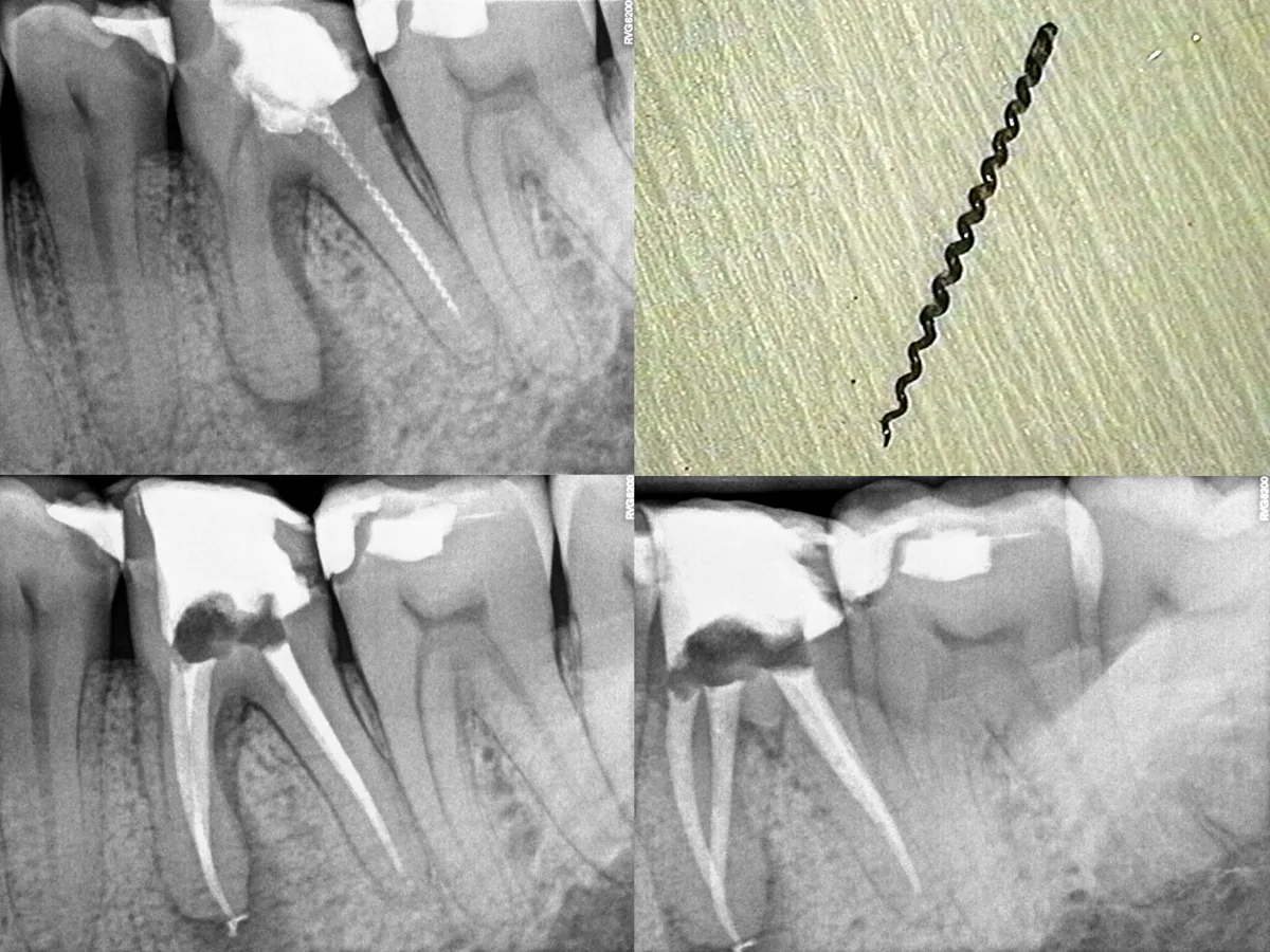 05Tooth 36
Removal of a Lentulo spiral from the canal
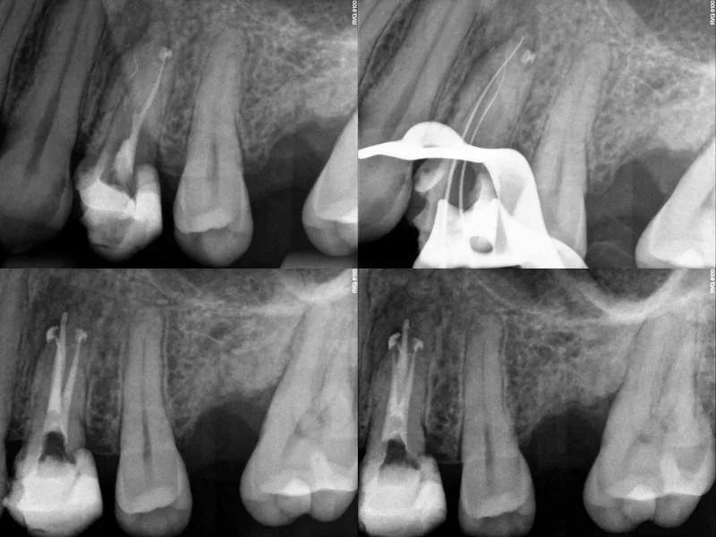 06Tooth 24
Three-canal premolar with complex anatomy
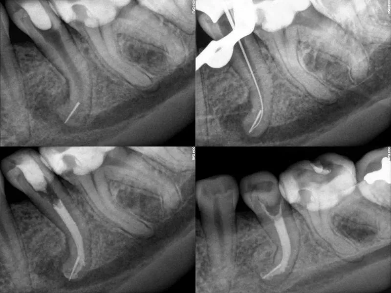 07Tooth 35
Bypassing a broken instrument and completing treatment
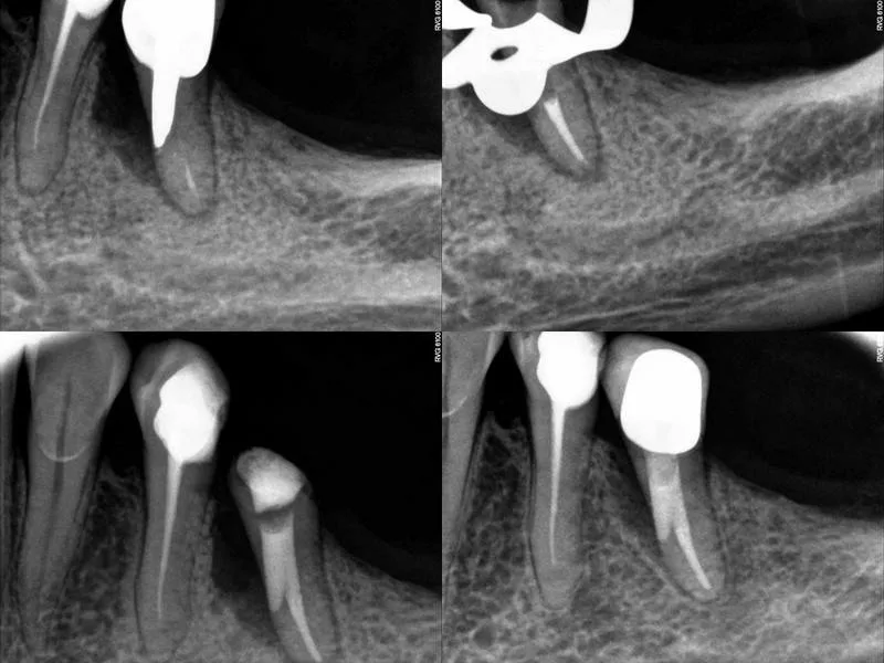 08Tooth 35
Post removal, perforation closure and retreatment
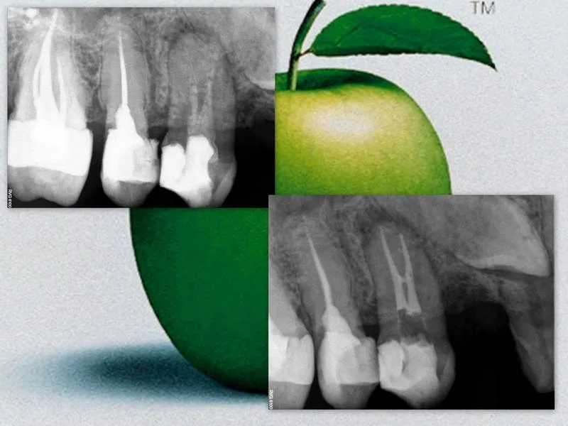 09Tooth 14
Microscope-guided root canal retreatment
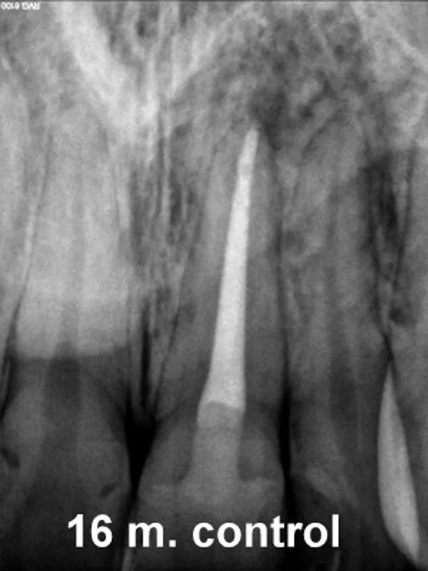 AFTER
AFTER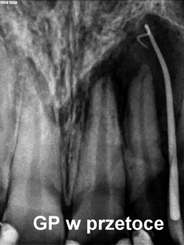 BEFORE
Large periapical lesion with a draining fistula — drag to see the healing
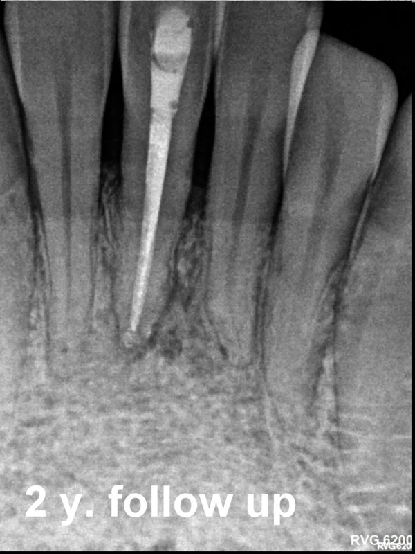 AFTER
AFTER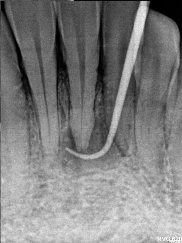 BEFORE
Retreatment of a tooth with a draining fistula — drag to see the healing
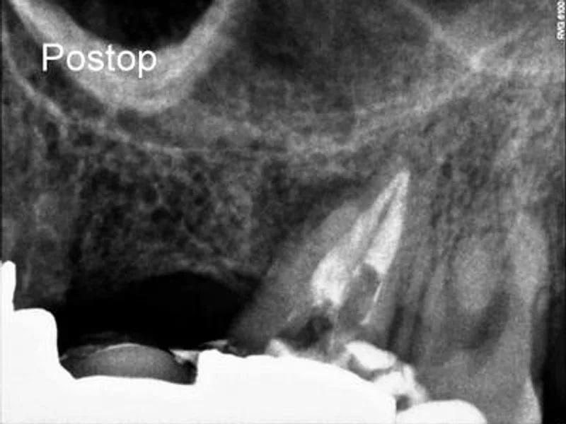 AFTER
AFTER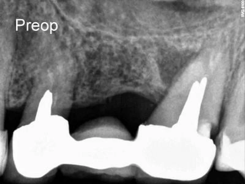 BEFORE
Retreatment under an existing bridge — drag to see the result
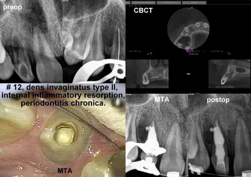 13
13Dens invaginatus with internal resorption
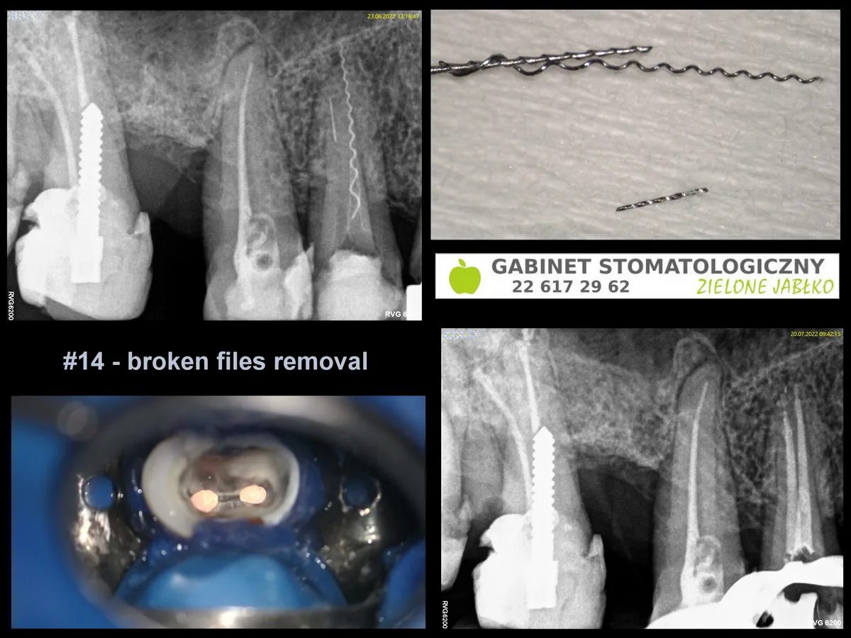 14Tooth 14
Fracture / difficult root anatomy case
[PLACEHOLDER — final selection sign-off from Dr Dziedzicka before go-live]
First-time treatment, first canal
each additional canal — 500 zł
1100 zł
Retreatment, first canal
each additional canal — 650 zł
1300 zł
Difficult case surcharge
curved canal, long root, difficult access
from 200 zł
Jagoda Chmiel
I wholeheartedly recommend it. Painless root canal treatment under the microscope, and very quick appointments too. Other clinics made me wait two months, but here the whole matter was resolved within a week. The doctor is very friendly and professional. She explains everything in detail. The office is spotless.
Wojciech Pykało
I rate the Doctor's work very highly. Attention to detail, commitment, and the equipment in the office (microscope, X-ray) guarantee success, especially in difficult cases (e.g. complicated root canal treatment that other dentists couldn't handle). My wife and I have been satisfied patients for several years. I recommend it!
Julia Czaban
The Doctor saved my tooth! I had to undergo repeat root canal treatment with a broken instrument — a task none of the four endodontists I had consulted would take on. They all agreed extraction and an implant was the only solution. Fortunately, on recommendation I found Dr Dziedzicka — and it was a breakthrough. She is a true specialist — with vast knowledge, precision and empathy. She devoted as much time to each visit as needed and explained the entire treatment in detail, showing the results on images. I am full of admiration and gratitude for the professionalism, commitment and care I received. I recommend her to everyone!!!